Tuesday, January 14, 2014

Pau Gasol's latest injury: strained flexor tendon

An MRI revealed that Pau Gasol has a moderately strained flexor tendon of his left big toe.  He injured it in the Lakers' 123-87 loss to the Clippers last Friday.


What does this injury mean for Pau Gasol?

The strained tendon is the flexor hallucis longus (FHL) tendon.  This tendon attaches to the base of the big toe and acts to flex the big toe.  In terms of the severity of the injury, it is fairly minor but it can affect how he plays.  The main function of the FHL tendon is to allow the big toe to push off.  It is important during running and jumping and helps to contribute to the lift a player gets.  If the flexor tendon is strained and not at 100%, Gasol will likely not be able to jump and run as well.

Tendon strains typically will take 2 to 3 weeks to heal but the return time can be improved with physical therapy.  The good news is that there was not any tear on the MRI.  Tendons do not have a great blood supply so tendon tears will typically require 6 to 8 weeks to heal with immobilization.  Occasionally tendon tears will not heal with immobilization and will require surgical repair of the tendon.  Pau Gasol should return without too much time missed and play at a similar level as he did before he had this injury.  

Thursday, January 9, 2014

Brook Lopez's foot surgery


Brook Lopez of the Brooklyn Nets suffered a broken 5th metatarsal in his right foot during a December 20 game against the Philadelphia Sixers.  He underwent season ending surgery on his foot last weekend and is expected to miss the rest of the season.  An additional procedure was done in addition to the repair of the fracture to help "unload and protect the injured area".  The theory is that it would redistribute the pressure sustained in that area more evenly.  This additional procedure was reportedly done to the first metatarsal.  Lopez has had issues with the 5th metatarsal before - injuring it in back in the 2011-12 season twice on the same right foot.

What does Brook Lopez have? 

Brook Lopez likely has a Jones fracture of the 5th metatarsal.  The 5th metatarsal is a long bone on the outside of the foot that consists of a head, neck, shaft and base.  Many nutrient arteries supply the long bone of the 5th metatarsal but there is one area where the shaft of the 5th metatarsal meets the base of the 5th metatarsal where there is poor vascular supply to the area.  Fractures that occur in this area of the 5th metatarsal - Jones fractures - are notoriously slow and difficult to heal due to this.

What is the procedure used to fix the Jones fracture?  

Jones fractures can be treated conservatively with casting and strict non-weightbearing if the bone is well aligned.  However, most will often require surgery to increase the probability of the bone healing.  Athletes in particular will often require surgery to speed up the healing process so that they can return to play quickly.  The surgery involves making sure that the fracture edges are fresh and bleeding so that new bone can form at the fracture site.  After this, a large screw is placed down the shaft of the 5th metatarsal to compress the fracture, giving it an ideal environment to mend and heal.  During this procedure, bone graft may added to the fracture site to help augment repair and promote healing.  After the surgery, the bone will often take 8 to 12 weeks to heal.  During that time, the patient is in a cast, non-weightbearing.

What is the additional procedure done to Brook Lopez? 

In addition to the fracture repair, reports have indicated that Lopez also underwent a separate procedure on the foot on the first metatarsal to help unload the injured area.  Jones fractures often will occur with increased stress to the fifth metatarsal.  Often, this is either due to "metatarsus adductus" or "pes cavus".  "Metatarsus adductus" is when the long bones in the foot point towards the midline of the body, creating a prominent 5th metatarsal and increased stress on the bone.  "Pes cavus" is when a high arch is present and this is often due to a dropped, or plantarflexed, first metatarsal.  When a plantarflexed first
metatarsal is present, the first metatarsal will often strike the ground first and the heel is unable evert/move to provide shock absorption.   This will often result in increased pressure on the outside of the foot where the 5th metatarsal is present.

The procedure that was likely done on Brook Lopez is an elevational first metatarsal osteotomy.  With the first metatarsal dropped causing increased pressure on the 5th metatarsal, elevating the first metatarsal in line with the other metatarsals should theoretically distribute the weight evenly across the foot and reduce the likelihood of another injury to that bone.  This procedure helps to correct "pes cavus".

Is this the right procedure? 

Brook Lopez has now had this injury several times and his foot type may be predisposing him to this injury.  Without examining him or seeing radiographs, it is hard to say whether or not it is the right procedure for him.  However, in theory, the additional procedure should offload the 5th metatarsal by allowing the heel to evert and put less pressure on the 5th metatarsal.  Less pressure means less stress on the bone and a lower probability of the bone breaking.  This procedure involves cutting a wedge in the bone and elevating the first metatarsal.  Screws are placed across the osteotomy site in order to hold the bone cut in place.

Since this is a recurrent injury and not an isolated one, it is valid to think that his foot type may be predisposing him to this injury.  If done correctly, I do believe that this procedure will effectively offload the 5th metatarsal and reduce his chance of injury in that same area.

Tuesday, October 8, 2013

Julio Jones' foot injury

There are reports that Julio Jones has sustained a season ending foot injury while playing last night in the Atlanta Falcons' 30-28 loss to the New York Jets.  According to reports, Jones continued to play through the injury.  Jones had a 5th metatarsal fracture in 2011 - the same foot that was injured last night.  A specific diagnosis has not been reported yet, as Jones is seeking a second opinion, but there has been speculation that the injury is a Lisfranc ligament tear.

What is the Lisfranc ligament and how is it injured? 


The Lisfranc ligament is a strong ligament in the midfoot that helps to stabilize the foot.  As pictured in the image above, it is attached from the medial cuneiform to the 2nd metatarsal base.  When this ligament is disrupted, there will be a displacement of the bones.  This displacement can lead to instability of the foot as well as degenerative joint changes (arthritis) in the future.  The ligament can be disrupted from a direct or indirect force on the foot.  An example of a direct force on the foot would be a heavy object dropping on the foot.  An indirect force on the ligament would be from a rotational force on the foot while the foot was pointed downwards.  

How is a Lisfranc injury diagnosed and treated?  

A Lisfranc injury is diagnosed clinically with the help of imaging studies such as x-rays and MRIs.  Lisfranc injuries can vary in severity - from a mild sprain to a tear in the ligament to fractured bones.  X-rays reveal if there is any displacement of the bones.  If there is no displacement but pain when pushing or stressing the Lisfranc ligament.  In these cases, an MRI may reveal a mild sprain of the ligament.  In these cases, the patient is casted and immobilized with a cast for about 6 weeks before progressing to a walking boot.  If x-rays show a displacement of the bones and/or fracture(s) of the midfoot, surgery is indicated in athletes, as even a small displacement of the bones can lead to long term disability and pain.  Surgery is done to realign the ligament and reduce any associated fractures that may be present in the foot.  Recovery after surgery requires 6 weeks in a non-weightbearing cast before transition to a walking boot for 2 weeks.  Typically an athlete will require a few months of rehab before returning to their sport depending on the severity of the injury.  

Does Julio Jones have a Lisfranc injury? 

With the limited information that we do know - it is possible that Julio Jones may have a Lisfranc injury.  Seeing as he was able to play through the injury is encouraging as it likely means it is a fairly mild injury.  However - he was able to play through a foot fracture in the past so his pain tolerance may be more than most.  With a average timeline of 3 to 4 months for return, it fits the timeline of him missing the rest of the season - which is what early reports indicate.  If he does have a Lisfranc injury, it is important to have the bones realigned if they are displaced to maintain stability of the bones in the midfoot.  Once they are realigned and the ligament is healed there should be minimal to no residual problems for him in the future.  It is possible that he may have a new fracture in his 5th metatarsal as well.  With a surgically repaired 5th metatarsal Jones fracture, a screw is often used to fix it.  The screw can occasionally serve as a stress riser and put the bone at risk for a stress fracture.  In fact, a CT scan is a clue that the problem is likely boney rather than involving soft tissues (like a Lisfranc ligament) unless the Lisfranc ligament tear had an associated fracture.

Update: Julio Jones does indeed have a fracture in the area of his previous injury and is scheduled to have surgery on Monday October 14th, 2013.  The procedure is likely to remove the previous hardware, clean up the fracture, and fix it again with screws and/or plates.  A period of 6-8 weeks immobilization is needed to allow for healing of this fracture.  Due to the lack of blood supply to the Jones fracture site, it may take longer.

Follow me on Twitter @sydneyyau


Monday, September 30, 2013

Why Matt Kemp is out for the playoffs

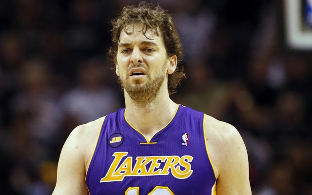
Matt Kemp, who is an outfielder for the Los Angeles Dodgers, will be unable to help the his team in the post season this year due to an MRI showing swelling in one of the major weightbearing bones in Matt Kemp’s ankle, the talus.  Kemp initially injured his ankle in a play at the plate against the Washington Nationals on July 21st.  After missing 52 games on the disabled list, he returned to play on September 16th.  However, he was held out of Saturday’s game due to soreness in his ankle.  This prompted an MRI that revealed swelling in the talus, taking Kemp out of the lineup in the post season. 

What does swelling in the ankle (talus) mean?

On an MRI after an ankle sprain injury, swelling in the talus typically will mean that there is some cartilage damage in the bone, called an osteochondral lesion.  When an ankle is sprained, a number of different structures can be injured.  In addition to the ligaments and tendons around the ankle being torn, occasionally a small piece of cartilage can chip off of the talus bone.  This cartilage damage is usually not readily seen on x-rays, but will often show up on MRI as bone swelling.  A CT scan may be useful in determining the size of the lesion. 

How did this happen? 

When there is an ankle sprain, many structures in the area can also be injured as well.  In addition to ligaments tearing, the peroneal tendons (tendons on the outside of the ankle) can stretch and tear as well.  Additionally, the ligament may tear off bone at its attachment site.  Depending on the severity of the sprain, the cartilage may chip off if there was any impact in the area.  According to reports, Matt Kemp's previous MRIs did not show any swelling of the bone.  Therefore, this injury may have been a result of a loose, or unstable ankle.  If the ligaments do not heal correctly, they may become loose.  Ligaments are strong structures that help to prevent unwanted motion.  When they are too loose, however, the ligaments may not be able to hold joints in place and there may be excess and unwanted motion at the joint.  Over time, there may be rubbing of the bone due to these loose ligaments which may cause cartilage damage or bone swelling in the ankle bone.  I suspect that the ligaments may be a little loose, causing a little movement in the ankle while Matt Kemp was playing.  This small movement may have been enough to cause irritation of the ankle and possibly a cartilaginous injury.   

What is the treatment of an osteochondral lesion?

Treatment of an osteochondral lesion depends on the location and severity of the lesion.  Lesions on the inside, or medial aspect, of the talus are usually deeper and more stable.  Lesions on the outside, or lateral aspect, of the talus are usually shallower and wafer shaped – making them less stable.  Osteochondral lesions can be classified as compression or bruising of the bone (stage 1), partially detached (stage 2), completely detached but non-displaced (stage 3), or completely detached and displaced in the ankle joint (stage 4).  Stage 1, 2, and lateral stage 3 lesions are best treated conservatively with a period of immobilization that includes a non-weightbearing below knee cast for six weeks.   If pain persists after this period of immobilization, surgery may be indicated.  Medial stage 3 and stage 4 lesions are best treated with surgery.  

What are the surgical options for osteochondral lesions?

There are a number of procedures that can be done depending on the size and depth of the lesion. Before surgery, the size and depth of the lesion should be determined with a CT scan to help with planning the appropriate procedure.

Microfracture surgery

Cartilage in general has a poor blood supply and it has poor healing potential.  Microfracture surgery involves drilling holes into the lesion to stimulate blood flow in the area and promote the formation of fibrocartilage.  This procedure is fairly minimally invasive as it is often done through a scope.  This procedure has good outcomes for smaller lesions.

Cartilage graft (OATS procedure) 

A cartilage graft can be placed in the area of the osteochondral defect to effectively replace damaged cartilage.  This method is usually reserved for lesions of about 1 cm in diameter.  In this procedure, the osteochondral lesion is punched out and replaced with a cartilage graft of identical size from a donor.  For the best results, a graft is taken from the similar bone in which the osteochondral defect is present as to recreate the anatomic contour of the joint as best as possible.  This is best done with a cadaver bone that matches the affected ankle.  Cadaver bones should be used within 14 days of it being harvested and should be fresh, not frozen.  Frozen grafts will deteriorate cartilage cells and reduce the ability of the graft to successfully incorporate into the host.

Stem cells

Mesenchymal stem cells make up about 2-3 % of all blood cells in bone marrow and they have the ability to differentiate into different types of cell types if placed in the right environment.  They also have the ability to stimulate new blood vessel growth, which is important in developing avascular tissue such as cartilage.  Stem cells can be separated from bone marrow that is harvested from the body and either injected into the ankle joint or placed over the osteochondral lesion itself in a gel form with a scaffold graft.  The stem cells will then differentiate into cartilage due to the growth factors and signals that are present in the environment in which they were placed.  

What should Matt Kemp do?

The current treatment plan for Matt Kemp is immobilization in a non-weightbearing cast.  This likely means that the lesion is either a stage 1, 2, or lateral stage 3 lesion and likely to heal with conservative treatment.  It is very important to rest this injury because the talus bone does not have a great blood supply.  Any additional movement or pressure to the lesion may prevent the lesion from healing.  Over time, an untreated osteochondral lesion can lead to ankle arthritis.  Therefore it is not an injury that he should play through, as it can cause further damage to the ankle.  A CT scan would be useful in evaluating any cartilage or bone defect that may be present in the ankle.  Should he continue to have pain and/or swelling to the ankle after his period of immobilization, surgery may be indicated.  After immobilization, a period of rehab is needed.  Range of motion and strengthening exercises are done to help strengthen the ankle.  Assuming conservative treatment is successful, Matt Kemp should be back for spring training in 2014 with minimal residual effects from the ankle injury.

Monday, July 29, 2013

Albert Pujols' out for the season?

Albert Pujols has been suffering from plantar fasciitis for 6 or 7 years and reports indicate that he may have torn his plantar fascia last Friday.  He was placed on the disabled list and will be getting an MRI.  This MRI would reveal whether or not the fascia is torn.

I discussed Pujols' condition in an earlier blog post and it appears that the fascia may have degenerated enough causing a tear.  Tearing of the fascia may actually be beneficial for Pujols as the tension may be relieved from the ligament pulling at the heel bone.  However, a tear in the fascia is painful and requires time to heal.  A tear in the fascia may sideline him for the rest of the year.

Plantar fascia tear

The plantar fascia is a strong ligament on the bottom of the foot that helps to support the arch.  It attaches at the heel and fans out to attach to each of the toes.


A plantar fascia partial or full tear can occur in any of the bands, but the medial band (the band closest to the big toe) is the most common.  

Cortisone injections, which are commonly used to treat plantar fasciitis, can be the source of a plantar fascia tear, as they can often weaken the integrity of the plantar fascia.  Studies describing the incidence of a plantar fascia rupture after injection range from 2.4-22% (1-3).  Degeneration of the fascia may also play a role in a plantar fascia tear.  Patients suffering with plantar fasciitis for long periods of time eventually develop plantar fasciosis.  In this state, the plantar fascia thickens and develops scar tissue.  This thickening can cause a loss of integrity to the fascia, increasing its risk of rupture.  

Albert Pujols' case

In Pujols' case, he has been suffering from this problem on and off for 6 to 7 years and likely has plantar fasciosis.  If he had been treated with corticosteroids - as many athletes are - his risk of developing a partial tear may have increased as well.  Should Pujols have a partial tear in his fascia, he needs to be immobilized and the fascia needs to be supported.  Depending on the severity of the tear, he may be out 1 to 2 months recovering.

References:
1. Kim C, et al.  Incidence of plantar fascia ruptures following corticosteroid injection.  Foot Ankle Spec 2010; 3(6):335-7
2. Acevedo JI and Beskin JL.  Complications of plantar fascia rupture associated with corticosteroid injection.  Foot Ankle Int 1998; 19(2):91-7
3. Saxena A and Fullem B.  Plantar fascia ruptures in athletes.  Am J Sports Med 2004; 32(3):662-5


Thursday, July 25, 2013

Tim Hudson's unfortunate injury

Sydney K Yau, DPM.  Follow me on Twitter @sydneyyau

Atlanta Braves pitcher Tim Hudson sustained an ankle fracture last night while covering 1st base.  The runner, Eric Young Jr. of the New York Mets, inadvertently stepped on Hudson's lower leg trying to get to 1st base ahead of the throw.  Hudson was immediately in pain and fell to the ground.  See the injury here:


Hudson is to undergo surgery when the swelling subsides.

Explaining the injury

Looking at the replay, you can tell that Young Jr. lands on Hudson's lower leg and causes the lower leg to flatten and turn out.  Hudson likely has a tibia and fibula fracture above the ankle joint and possibly a syndesmotic injury.  The tibia and fibula are the leg bones of the body.  Together they connect the knee to the ankle and form the ankle joint.  They are held together near the ankle by a strong ligament called the syndesmotic ligament or interosseous membrane.  This ligament provides stability to the ankle and is important for many athletes.  Occasionally this ligament is sprained and is diagnosed as a "high ankle sprain".


This fracture is a result of direct impact so it does not follow any typical fracture patterns from ankle fractures sustained from indirect impact such as from twisting injuries.  Looking at the replay, it appears that Young Jr. lands above the ankle joint at the lower leg.  This is important as it does not appear that the fracture affects the ankle joint itself.  If the fracture was within the ankle joint, he is at risk for arthritis in the joint and this may delay his healing and affect his rehabilitation.  If the fracture, as I suspect, is above the ankle joint, the tibia and fibula can be realigned with surgery and there is less risk of arthritis in his joint.  If there is separation of the tibia and fibula on radiographs, this means that the syndesmosis is torn and needs to be repaired as well.

Fixing the injury

Surgery is needed to realign the tibia and fibula and possibly repair the syndesmotic ligament as well. The fracture will likely be reduced surgically and held together using plates and screws.  If the syndesmotic ligament is torn, screws are often placed across the ligament to repair the ligament.  The surgeon should check the alignment of the bones in all three planes and make sure the ankle joint is in anatomic position to reduce risk of arthritis in the future.  This is especially important should the fracture extend into the ankle joint.  The surgeon should also check to make sure adequate length is maintained of the bone to prevent shortening, causing a limb length discrepancy.  This is more of a risk if the fracture is in multiple pieces, which may be possible in this case.

Hudson's prognosis

I believe that Hudson is likely done for the season.  Recovery time for the bones to heal often will last 6 to 8 weeks.  After the bones heal, Hudson will need to rehab his ankle for at least a month or two to restrengthen his leg and redevelop stability in his ankle should the syndesmotic ligament be torn.  The biggest risk after surgery is arthritis in the ankle and instability of the ankle.  This can happen if the ankle is not well aligned after surgery.  Due to the healing times, I believe he will likely be unable to start pitching until after the current season ends.  However, as long as the fracture is reduced back into its correct anatomic position and bone is allowed to heal, I believe this shouldn't be a career ending injury for Hudson and he should be back pitching next season for the Atlanta Braves unless he chooses to retire.

Tuesday, July 23, 2013

Matt Kemp returns! (and injures himself again).

Matt Kemp of the Los Angeles Dodgers returned to the lineup 2 nights ago and had 3 hits, including a home run, to help the Dodgers to a 9-2 victory.  However, he was taken out of the game in the ninth inning on a play at home plate.


On the night of the injury, Dodger manager Don Mattingly said that the injury appears to be a sprain and that no x-rays were planned to be taken.  He did not anticipate that Kemp would need to go to the disabled list.  However, last night, Don Mattingly was less clear on this issue.  He said that Kemp would likely miss the 3 game series against the Blue Jays and that Kemp actually may head back to the disabled list for the 3rd time this season.

Ankle Sprains

The ankle has many ligaments that holds the joint together and makes it more stable.  They are slightly elastic but have a firm end point that can be reached to prevent abnormal motion from occurring in the joint.  An ankle sprain occurs when the force exerted on the ligaments exceeds its failure point.  The ligament can tear or rupture depending on the extent of the force that is applied to it.  If the bone is actually weaker than the ligament, a piece of bone can be avulsed off by the ligament.

Matt Kemp's injury
As seen in the gif above, Matt Kemp sustained an inversion type ankle sprain.  In this sprain, the ligaments that are affected are on the outside of the ankle.  There are 3 ligaments on the outside of the ankle that prevent excess inversion of the ankle.  These are the anterior talo-fibular ligament (ATFL), calcaneal-fibular ligament (CFL) and posterior talo-fibular ligament (PTFL).  When these ligaments get sprained, they can stretch or even tear.  Judging from his injury and the amount of inversion that the ankle went through, it is likely that the ligaments tore.

Matt Kemp's outlook? 

According to reports, no x-rays were planned.  This is confusing to me; especially since he was seen limping in the locker room afterwards.  If there is an avulsion fracture, this may take longer to heal and recuperate from.  The bone needs to heal to maintain stability of the ankle.  X-rays are inexpensive and very non-invasive and there is little to no harm to take x-rays to evaluate the ankle after an injury like this.

Having said this, if this is a a ligament sprain is fairly easy to treat but it requires time.  Athletes may take 2-6 weeks to recover from this depending on the severity of the sprain.  Treatment requires immobilization, bracing, and physical therapy.

During treatment of a sprain, it is important to immobilize the ankle so that the ligaments can heal in its natural, anatomic position.  If the ligaments heal in a stretched out position, the ankle sprain may result from an unstable ankle and may eventually lead to arthritis.

I think Matt Kemp will recover well from this injury but it may take at least 2-3 weeks for him to feel stable on his ankle.  If he has an avulsion fracture, this may take 6-8 weeks for him to come back.